New Method: Could Colonoscopy Become a Less Invasive Examination?
For decades, colonoscopy has been one of the most important examinations used to look inside the large intestine. It can help doctors detect polyps, abnormal tissue, bleeding, inflammation, and colorectal cancer. It can also allow doctors to remove certain polyps during the same procedure.
However, traditional colonoscopy is an invasive examination. It requires a flexible tube with a camera to be carefully inserted through the rectum and guided through the colon. Although the procedure is generally considered safe, many people find the preparation and examination uncomfortable or unpleasant.
That is why researchers have spent years developing alternative ways to examine the colon.
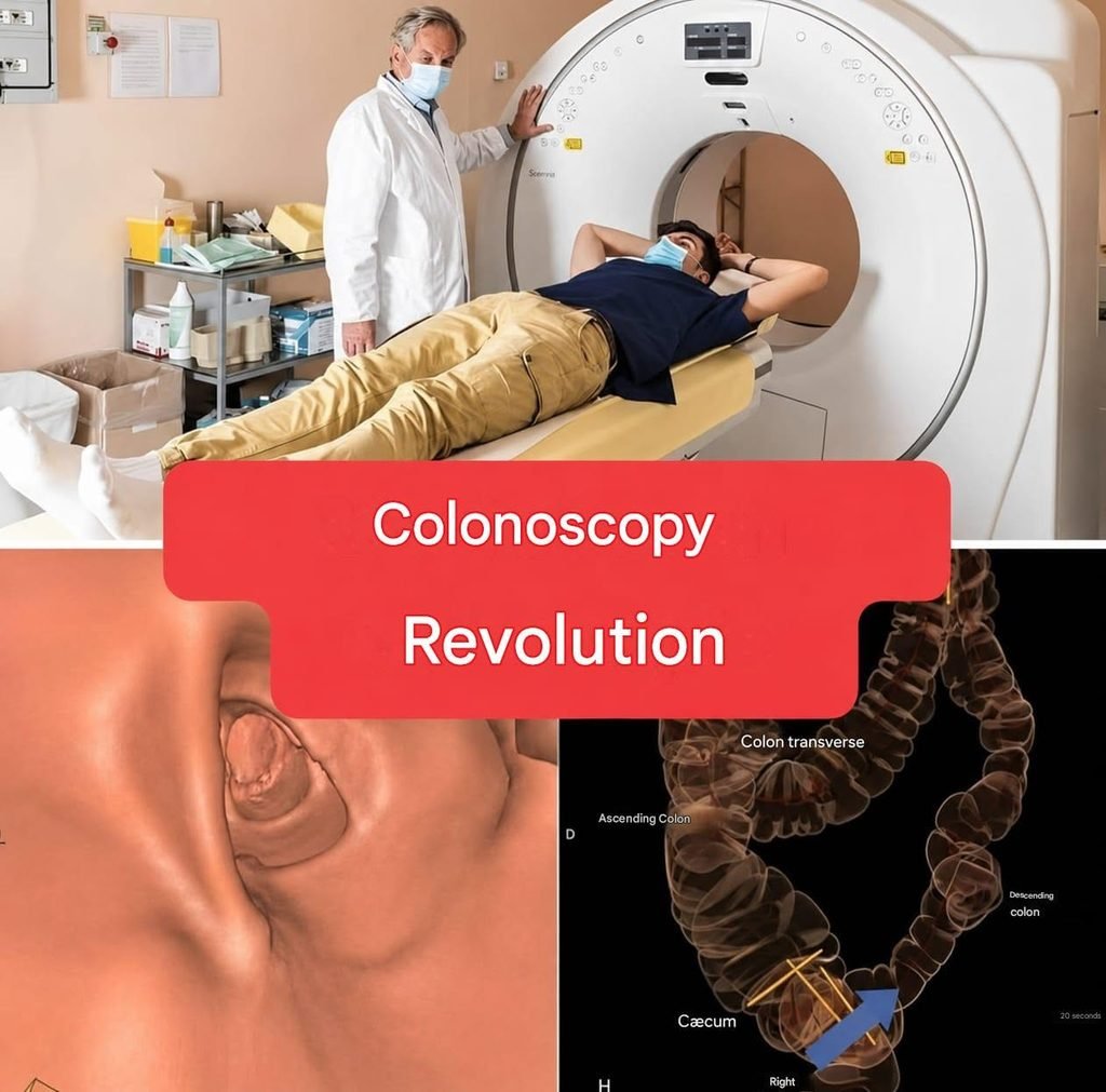
One of the most interesting developments is CT colonography, sometimes called a virtual colonoscopy. It uses computed tomography imaging to create detailed images of the colon without inserting a conventional flexible camera throughout the intestine.
But does this really mean that traditional colonoscopy is becoming obsolete?
The answer is more complicated.
What Is a Traditional Colonoscopy?
A conventional colonoscopy is performed using a long, flexible instrument called a colonoscope.
The device contains a small camera and light. During the examination, a trained medical professional guides the instrument through the rectum and into the colon.
The camera sends images to a monitor, allowing the medical team to examine the lining of the large intestine.
One major advantage is that colonoscopy is not only a diagnostic examination.
If a doctor finds a polyp, for example, it may be possible to remove it during the same procedure. Tissue can also be collected for laboratory examination when necessary.
This combination of seeing, sampling, and treating is one reason conventional colonoscopy remains so important.
Why Are Researchers Looking for Alternatives?
Although colonoscopy is highly useful, it has several disadvantages.
The bowel generally needs to be thoroughly cleaned beforehand. This usually involves a bowel-preparation solution that causes repeated bowel movements.
For many people, the preparation is actually one of the most unpleasant parts of the entire process.
The examination itself can also be uncomfortable. Depending on the circumstances, sedation may be offered or recommended.
There are also small risks associated with invasive procedures, including bleeding and, very rarely, perforation of the colon.
Because colorectal cancer screening is so important, researchers have looked for methods that could make screening easier and more acceptable to more people.
The goal is not simply to make the procedure more comfortable.
A major goal is to make screening accessible to people who might otherwise avoid it.
What Is a Virtual Colonoscopy?
CT colonography is an imaging-based examination.
Instead of guiding a camera through the entire colon, a CT scanner produces a series of detailed images of the abdomen and pelvis.
Special computer software can then reconstruct these images to create views of the inside of the colon.
In simple terms, doctors can examine the colon using images generated from CT scanning rather than relying entirely on a physical camera traveling through the intestine.
This is why the procedure is sometimes described as a virtual colonoscopy.
The patient does not have a conventional colonoscope traveling through the entire colon.
Is It Completely Non-Invasive?
This is where headlines can sometimes become misleading.
Virtual colonoscopy is less invasive than conventional colonoscopy, but it is not necessarily completely non-invasive.
The colon still generally needs to be cleaned before the examination.
A small tube may also be placed into the rectum so that the colon can be gently inflated with air or carbon dioxide. This allows the walls of the colon to be separated and makes abnormalities easier to see on the CT images.
Therefore, saying that there is absolutely “no invasion” would be an oversimplification.
The examination avoids the passage of a long camera through the colon, but it still involves preparation and some physical intervention.
What Happens During the Examination?
The process is generally much shorter than many people imagine.
After the bowel has been prepared, the patient lies on the CT scanner table.
A small tube is placed into the rectum.
The colon is gently expanded with air or carbon dioxide.
The patient then passes through the CT scanner while images are taken.
The scanner collects multiple images from different angles.
Computer software subsequently processes those images to produce detailed representations of the colon.
The patient may be asked to change position during the scan so that different areas can be visualized properly.
Unlike a conventional colonoscopy, there is no long flexible camera traveling through the entire colon.